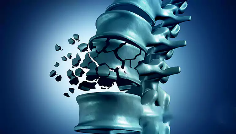
Spinal trauma and fractures encompass injuries to the thoracic or lumbar spine—ranging from minor stable fractures to severe unstable trauma. These can arise from accidents, falls, sports injuries, or underlying bone conditions, and may affect spinal stability, neurological function, and quality of life.
Symptoms may include: acute back pain, possible nerve-related symptoms (numbness, weakness), or neurological signs due to compression or instability. Severe trauma may also involve deformity or spinal cord injury. Minor fractures (isolated spinous or transverse process, laminar injury) often remain stable and neurologically intact.
Diagnosis should incorporate:
CT scans for precise fracture characterization
MRI to assess spinal cord and soft tissue involvement
Grading using AO‑spine classification and Morphological Modifiers like canal compromise or instability, which guide management decisions.
Appropriate for stable fractures without neurological compromise, such as compression-type fractures or non‑displaced burst fractures.
Rapid mobilization—with effective analgesia to facilitate early movement and prevent complications of immobility.
Pain management—NSAIDs, paracetamol, opioids (carefully in elderly), supportive analgesics guided by comorbidities.
Physiotherapy—tailored core stabilization, posture correction, and gradual ambulation under supervision.
Follow‑up imaging—standing radiographs after mobilization to ensure no deterioration; serial reevaluation within a week or more as needed.
Bracing—currently there is no consistent benefit in randomized trials; its use is case-dependent and not routine.
Conservative care requires close interdisciplinary coordination and adjustment if clinical or radiological deterioration emerges.
Surgery is warranted when:
Neurological deficits—especially with canal compromise
Unstable fractures—AO Type B or C injuries, dislocations, or burst fractures with kyphotic deformity
Significant canal stenosis from displaced bone fragments
Multiple injuries or compromised healing environment (e.g., poor bone quality or polytrauma)
Key surgical goals:
Prompt decompression to relieve pressure on neural elements
Stabilization via instrumentation—posterior, anterior, or combined fusion or fixation
Alignment restoration, especially addressing sagittal balance and preventing late kyphotic deformity
Use of minimally invasive or robotic-assisted techniques when feasible to reduce morbidity and enhance early mobilization
Kyphoplasty or vertebroplasty may be considered for compression fractures due to osteoporosis, offering rapid pain relief—particularly kyphoplasty (restores vertebral height)—though suitability depends on fracture type and bone quality.
The motto “Time is spine” underscores the importance of early decompressive surgery—ideally within 8–24 hours—for incomplete spinal cord injuries in thoracic or thoracolumbar segments, to reduce secondary injury and improve neurological outcomes .
Early decompression, stabilization, and correction enable improved spinal cord perfusion and better functional recovery.
A. Comprehensive Assessment
Detailed neurological exam, advanced imaging (CT + MRI), AO spine classification and morphology scoring.
Consideration of overall health, bone density, comorbidities, and patient activity demands.
B. Tailored Treatment Pathway
Stable, neurologically intact fractures → non-surgical management focusing on pain relief, mobilization, and rehab.
Unstable fracture types, neurological compression, deformity, or failed conservative care → surgical decompression and stabilization (favoring minimally invasive or robot-assisted techniques when available).
C. Precision Surgical Strategy
Posterior fixation and possible fusion for unstable injuries, with short-segment constructs when possible at thoracolumbar regions.
Anterior reconstruction or combined approaches for severe burst injuries or poor bone quality. Consider PMMA cement augmentation in osteoporotic bone under select cases.
D. Postoperative Care & Rehabilitation
Multimodal analgesia, early ambulation, tailored physiotherapy, gradual core strengthening.
Periodic imaging and clinical evaluation; plan hardware removal (if posterior only) within 6–12 months in stable, healed cases .