Spinal Infection - Tuberculosis

Service Details:

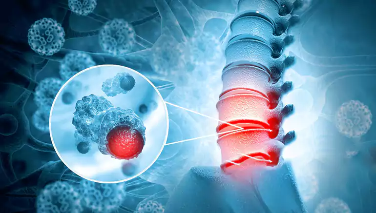
Spinal infection caused by Mycobacterium tuberculosis, leading to vertebral destruction, pain, and potential spinal deformity (Pott's disease). Data tracks inflammatory markers, imaging (characteristic vertebral changes), and response to anti-TB drugs.

Spinal tuberculosis, also known as Pott's disease or tuberculous spondylitis, is a destructive infection of the spine caused by Mycobacterium tuberculosis, the same bacterium that causes pulmonary tuberculosis. It typically spreads to the spine hematogenously (through the bloodstream) from a primary TB infection in the lungs or other parts of the body. The infection usually starts in the vertebral body, often affecting the lower thoracic and upper lumbar spine, and can then spread to adjacent vertebrae, intervertebral discs, and surrounding soft tissues, potentially forming abscesses (cold abscesses) that may track along muscle planes.
  • Data Collection:

    • Initial Diagnosis:

      • Clinical Presentation: Data includes a history of back pain (often chronic and progressive), stiffness, muscle spasms, possible neurological symptoms (weakness, numbness), low-grade fever, night sweats, weight loss, and a history of exposure to TB or a past TB infection.
      • Laboratory Tests: Data includes elevated inflammatory markers (ESR, CRP), a positive Mantoux test (tuberculin skin test) or Interferon-Gamma Release Assay (IGRA), and sputum or other extra-spinal samples positive for M. tuberculosis. A definitive diagnosis often requires a spinal biopsy (image-guided or surgical) to identify M. tuberculosis through culture or PCR and for histological examination showing characteristic granulomatous inflammation with caseous necrosis.
      • Imaging: X-rays may show characteristic findings like vertebral body destruction (lytic lesions), disc space narrowing, vertebral collapse leading to kyphosis (gibbus deformity), and paravertebral soft tissue swelling. CT scans provide more detailed bony architecture and can show calcifications within abscesses. MRI is crucial for visualizing soft tissue involvement, including epidural or paraspinal abscesses and spinal cord compression.
    • Short-Term Monitoring (During Treatment):

      • Clinical Response: Data tracks the reduction in pain (using pain scales), improvement in neurological function, resolution of fever and systemic symptoms, and any changes in spinal deformity.
      • Laboratory Markers: Serial measurements of ESR and CRP are used to monitor the inflammatory response to anti-tuberculous drugs.
      • Imaging: Follow-up MRI scans may be performed to assess the size and resolution of abscesses and to monitor for any progression of vertebral destruction or spinal cord compression.
    • Long-Term Monitoring (Post-Treatment):

      • Radiological Outcomes: Data includes monitoring the stability of the spine, the fusion of affected vertebral segments (spontaneous or surgically assisted), and the progression of any spinal deformity (kyphosis). Serial X-rays are often used for this.
      • Functional Outcomes: Assessment of the patient's ability to perform activities of daily living, pain levels, and neurological function using standardized scales (e.g., Oswestry Disability Index, Nurick grading for spinal cord compression).
      • Relapse: Monitoring for any recurrence of symptoms or radiographic evidence of active infection.
      • Complications: Data on long-term complications such as chronic pain, spinal instability, and neurological deficits.